
Diagnostic Ultrasound for Physio: A Buyer Checklist
Buying diagnostic ultrasound for a physiotherapy centre? Compare clinical scope, probes, operator training, records, cleaning and service support.
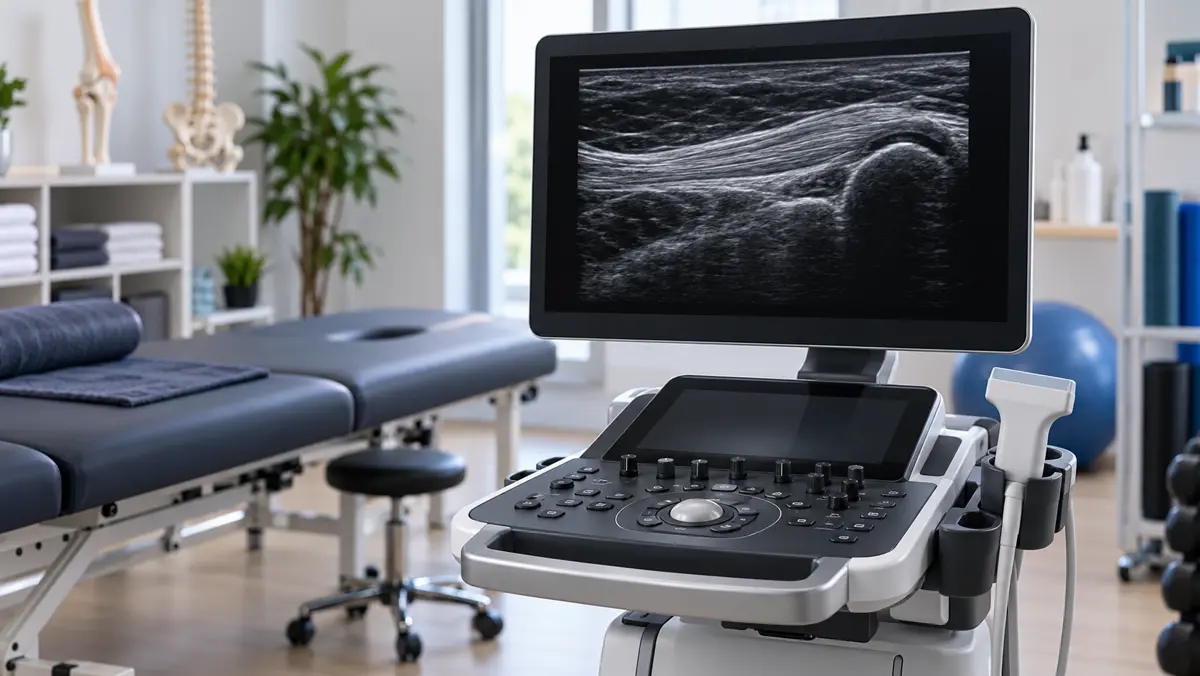
Diagnostic ultrasound is becoming more visible in sports medicine and rehabilitation settings. A high-quality image of a tendon or muscle can be useful when the clinical question and operator skill are right. But buying a scanner does not automatically create a diagnostic service. For a physiotherapy centre, the decision must include intended use, who is qualified to scan and interpret, what will be documented, how patients will be referred, and whether the equipment is suitable for the structures being examined.
This guide is a practical buyer checklist for diagnostic musculoskeletal ultrasound in a physiotherapy or rehabilitation setting. It distinguishes imaging from therapeutic ultrasound, explains the core specification questions, and places training, quality assurance and patient safety alongside the machine itself. It does not endorse a model or imply that every centre should offer diagnostic imaging.
Diagnostic and therapeutic ultrasound are different
Diagnostic ultrasound uses sound waves to create images of tissues. It can show selected muscles, tendons, ligaments, nerves and joints in real time. Therapeutic ultrasound uses sound energy as a treatment modality and does not produce a diagnostic image for interpretation. The machines, transducers, staff competencies and patient pathways are different.
This distinction matters because a supplier may use the single word "ultrasound" for both. Our shockwave versus therapeutic ultrasound guide covers treatment technologies. The present article is about an imaging system and the professional workflow needed to use it appropriately. A physiotherapy centre should not purchase a therapeutic device expecting it to answer imaging questions.
Diagnostic ultrasound also does not replace every MRI or X-ray. It is particularly operator-dependent and has limitations for deeper structures and some bone or joint questions. Ask what exact clinical decisions your centre expects it to support, and whether a qualified referral pathway is available for questions it cannot answer.
Define the clinical use before comparing brochures
List the body areas and questions you expect to assess. Are clinicians examining superficial tendons, peripheral nerves, muscles, joint effusions, or guiding a procedure? These are not identical use cases. A system that looks attractive in a sales demonstration may be poorly matched to the actual depth, field of view or workflow of your patients.
The American Institute of Ultrasound in Medicine's musculoskeletal practice parameters describe standards for performing and recording high-quality examinations. They emphasise that a complete examination requires more than obtaining one appealing image. Before buying, decide who is authorised and trained to perform which examinations in your jurisdiction, who interprets them, and how findings enter the patient's care plan.
Regulatory and professional scope rules vary. A US practice parameter is useful for quality thinking, but it is not an Indian licence or permission to provide a particular service. Verify local requirements with qualified advisers before marketing diagnostic scans.
Which probes and image features matter?
For superficial musculoskeletal structures, a linear-array transducer is commonly useful because it offers high-frequency imaging with good detail near the skin. Deeper structures may call for different probe characteristics. Ask a knowledgeable operator to test the exact system on representative examination types. A specification sheet's frequency range is not a substitute for seeing image quality in the hands of your team.
Compare depth penetration, near-field detail, frame rate for dynamic assessment, image optimisation controls, Doppler options where relevant, measurement tools and ease of saving labelled images. Consider whether the machine remains usable when the room is bright, the operator is wearing gloves, and the patient must change position. The best interface is one that supports consistent examinations, not the one with the longest feature list.
Portable scanners can save space and support movement between rooms, but battery life, screen size, probe protection and data transfer become important. Cart systems may offer a different ergonomic and workflow balance. Make a side-by-side demonstration checklist instead of deciding from product photographs.
Training is a core part of the purchase
Ultrasound images can look convincing even when the probe angle or scanning technique creates an artefact. Operators need anatomical knowledge, examination technique, recognition of normal variants and understanding of the method's limits. A new device does not give a practitioner the competence to diagnose every injury.
The AIUM diagnostic musculoskeletal accreditation resources connect diagnostic practice with training, documentation, cleaning and equipment quality assurance. Ask the supplier what training is included, who provides it, how competence will be assessed, and whether continuing education is available. Plan for new staff and periodic reassessment, not just one installation-day demonstration.
Define escalation rules. If an image is uncertain, who reviews it? When should the patient be referred for MRI, X-ray, specialist assessment or urgent care? A responsible service is willing to say "this scan does not settle the question." That limit protects both patients and professional credibility.
Documentation and data handling
A useful diagnostic examination should record the question asked, body side and area, relevant technique, representative images, findings and limitations. Images must be stored and retrievable according to the facility's privacy and record policies. A live image shown on a screen without a reliable report or record can be difficult to use in follow-up care.
Ask whether the scanner exports standard image formats, integrates with existing systems, supports secure user access and has a clear backup pathway. Check how software updates affect stored studies. If a third-party cloud service is involved, understand where data goes and who controls access. These operational questions are as important as probe specifications.
Our guide to 3D movement analysis for rehab centres discusses another assessment technology. In both cases, a measurement creates value only when it is reliable, interpreted appropriately and connected to a decision.
Cleaning, ergonomics and quality assurance
Transducers contact patients and must be cleaned according to their use and the manufacturer's instructions. Ask what cleaning products are compatible with the probe, how long the process takes between patients and whether replacement accessories are available. Inadequate cleaning can turn an attractive service into an avoidable safety risk.
Ultrasound work can strain shoulders, wrists and neck when room layout is poor. Test the height and movement of the screen, probe cable length, bed access and how the operator can remain close to the patient without awkward postures. Think about left- and right-sided examinations and wheelchair access. The AIUM resource library includes guidance on cleaning, quality assurance and operator ergonomics.
Quality assurance should include periodic checks that the probe and image performance remain suitable. A dropped transducer or faulty cable can degrade an image subtly. Staff should know how to report problems and pause use until the device is checked.
Supplier and service questions
Request the exact model and probe configuration in writing. Ask for installation requirements, training scope, warranty terms, software support, preventive maintenance, repair response and availability of replacement probes. Confirm whether the demonstration system matches the quoted system. A feature shown on a demo unit may require an additional module not included in your configuration.
The service plan should describe what happens when the device fails, not just the interval for a routine visit. Where are parts held? Is there a loan device? Who handles software issues? A scanner unavailable for weeks disrupts the patient pathway and staff confidence. Avoid relying on promises about guaranteed revenue; demand and operational fit differ between centres.
Our force plate versus isokinetic testing comparison makes a similar point: equipment should be selected for the specific assessment question, not because several devices share a broad "rehab technology" label.
A decision checklist for your centre
Before signing, confirm five things. First, the intended examination list is clear and clinically relevant. Second, appropriately qualified people will perform and interpret each scan within local rules. Third, the selected probes and software have been tested on real examples. Fourth, documentation, storage, cleaning, quality assurance and referral processes are written down. Fifth, the supplier can support the exact configuration after installation.
If one of these is unresolved, pause the purchase and close the gap. A centre may decide that a referral relationship with an imaging provider is more appropriate than owning a machine at its current stage. That can be a professional decision, not a missed opportunity. If your team is evaluating options, contact Regenis with your intended use and facility workflow.
The takeaway
Diagnostic musculoskeletal ultrasound can be valuable when trained operators use the right equipment for a defined question and document the result properly. It is not therapeutic ultrasound and not a universal replacement for other imaging. The strongest purchasing decision joins clinical scope, image quality, training, records, hygiene and service into one plan.
Frequently asked questions
Is diagnostic ultrasound the same as physiotherapy ultrasound?
No. Diagnostic ultrasound creates images for assessment. Therapeutic ultrasound applies sound energy as a treatment and does not provide the same diagnostic imaging workflow.
Which probe is commonly used for superficial tendons?
A high-frequency linear-array probe is commonly useful for superficial musculoskeletal structures. The exact choice depends on depth and the examination, so test it with a qualified operator.
Can any physiotherapist diagnose an injury with a new scanner?
No. Equipment ownership does not establish training, competence or legal scope. Verify local rules and ensure qualified interpretation and referral pathways.
Can ultrasound replace MRI for every sports injury?
No. It is useful for selected questions but has limits. The clinician should know when MRI, X-ray or specialist assessment is more appropriate.
What records should a scan create?
The service should record the clinical question, body area, representative images, findings, limitations and an accessible report under the facility's privacy procedures.
What should be checked before buying a used machine?
Verify probe condition, image quality, software and data access, service history, compatible cleaning instructions and availability of parts. Have a qualified operator assess performance before accepting it.
Sources and scope
This guide draws on AIUM musculoskeletal practice parameters, diagnostic MSK ultrasound accreditation resources and the AIUM resource library. It is equipment-planning education, not a statement of local legal scope or a substitute for professional training.